Abdominal Blood Vessels Labeled / Dissection of the blood vessels posterior to the diaphragm procedure:
byAdmin•
0
Abdominal Blood Vessels Labeled / Dissection of the blood vessels posterior to the diaphragm procedure:. In human anatomy, inferior epigastric artery refers to the artery that arises from the external iliac artery.it anastomoses with the superior epigastric artery.along its course, it is accompanied by a similarly named vein, the inferior epigastric vein.these epigastric vessels form the lateral border of hesselbach's triangle, which outlines the area through which direct inguinal hernias protrude. Microdissection of the artery, its main branches, and the perforator vessels was undertaken in 20 cadavers. Efferent branchial arteries injected with red latex. Advertising on our site helps support our mission. As the abdomen and pelvis contain the majority of internal organs, these regions need to be supplied by an extensive network of arteries and veins.
The aorta is the large artery leaving the heart. The aorta is the largest blood vessel in the body. This artery is responsible for transporting oxygen rich blood from your heart to the rest of your body. The superior vena cava is the large vein that brings blood from the head and arms to the heart, and the inferior vena cava brings blood from the abdomen and legs into the heart. This video series covers the blood vessels for anatomy and physiology ii students.
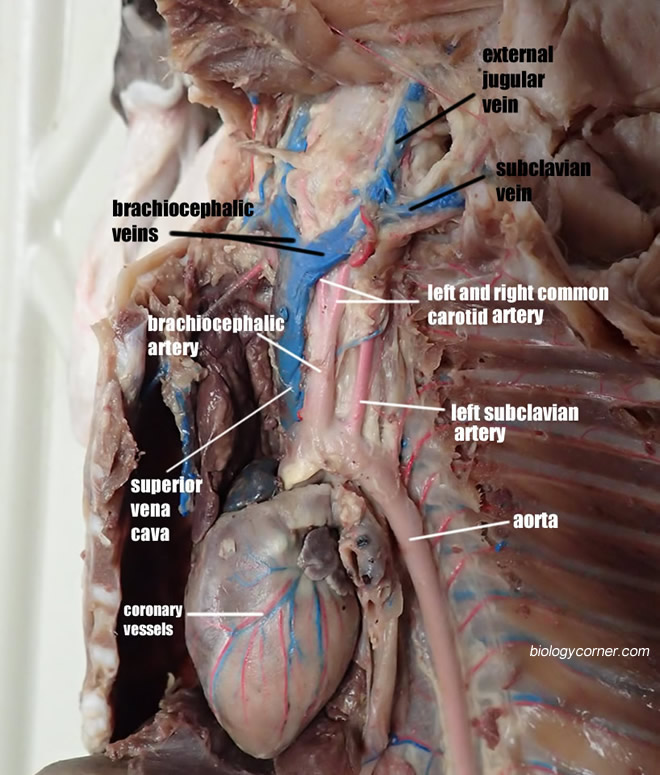
Cat Vessels Image Gallery from www.biologycorner.com I hope this anatomy guide is helpful. The presence of vascular loops allows surgeons to ligate individual vessels with the expectation that blood will find its way to a particular region by alternate branches. Practice identifying the blood vessels on the photographs here and in your fetal pig photoalbum online. The abdominal aorta enters the abdomen through the diaphragm at the level of the twelfth thoracic vertebre and continues to just below the umbilical area, where it splits into the right and left common iliac arteries. Structure of blood vessel walls. Instant anatomy is a specialised web site for you to learn all about human anatomy of the body with diagrams, podcasts and revision questions Physiology and anatomy of blood vessels prepared by dr. The suprarenal abdominal or paravisceral segment, inferior to the diaphragm but superior to the renal arteries.
In human anatomy, inferior epigastric artery refers to the artery that arises from the external iliac artery.it anastomoses with the superior epigastric artery.along its course, it is accompanied by a similarly named vein, the inferior epigastric vein.these epigastric vessels form the lateral border of hesselbach's triangle, which outlines the area through which direct inguinal hernias protrude.
The artery was found to be associated with two veins in most of the cases (90 percent). Practice identifying the blood vessels on the photographs here and in your fetal pig photoalbum online. 44 44shareswelcome to our series of articles on small animal abdominal ultrasonography. Finally, through ct scans the major blood vessels of the abdomen can be examined. Of course, recognition of the normal vascular anatomy is essential for the investigation of any abdominal vascular problem. I hope this anatomy guide is helpful. Label the intestinal structures using the hints provided. The initial articles provided an overview of basic ultrasonography principles and a discussion about how to perform a systematic scan of the abdomen. As the abdomen and pelvis contain the majority of internal organs, these regions need to be supplied by an extensive network of arteries and veins. The identification of abdominal vessels using ultrasound is based on knowledge of their normal location, appearance and relationship to specific organs. The infrarenal segment, inferior to the renal arteries and superior to the iliac bifurcation. This artery is responsible for transporting oxygen rich blood from your heart to the rest of your body. Read the other small animal abdominal ultrasonography articles.
As the abdomen and pelvis contain the majority of internal organs, these regions need to be supplied by an extensive network of arteries and veins. The initial articles provided an overview of basic ultrasonography principles and a discussion about how to perform a systematic scan of the abdomen. The common iliac arteries and veins. In human anatomy, inferior epigastric artery refers to the artery that arises from the external iliac artery.it anastomoses with the superior epigastric artery.along its course, it is accompanied by a similarly named vein, the inferior epigastric vein.these epigastric vessels form the lateral border of hesselbach's triangle, which outlines the area through which direct inguinal hernias protrude. Learn vocabulary, terms, and more with flashcards, games, and other study tools.
Flat Wire Model from classroom.sdmesa.edu Efferent branchial arteries injected with red latex. The aorta is the largest blood vessel in the body. The aorta is the large artery leaving the heart. The abdominal aorta supplies blood to much of the abdominal cavity. Label the abdominal contents using the hints provided. Doppler studies of the abdominal vessels demand an understanding of normal and abnormal blood flow patterns. The identification of abdominal vessels using ultrasound is based on knowledge of their normal location, appearance and relationship to specific organs. Read the other small animal abdominal ultrasonography articles.
Nerves, blood vessels, and lymphatics are present throughout.
Abdominal wall anatomy that is clinically pertinent to the surgeon, focusing primarily on the structures of the anterior abdominal wall, will be reviewed. The abdominal aorta is the largest blood vessel in the abdomen. Structure of blood vessel walls. Nerves, blood vessels, and lymphatics are present throughout. Read the other small animal abdominal ultrasonography articles. Common incisions and closure techniques, and prevention and management of wound complications, are discussed elsewhere. Instant anatomy is a specialised web site for you to learn all about human anatomy of the body with diagrams, podcasts and revision questions The common iliac arteries and veins. • they are also common in abdominal organs, the heart, and the brain dr. The vasculature is a network of blood vessels connecting the heart with all other organs and tissues in the body. Doppler studies of the abdominal vessels demand an understanding of normal and abnormal blood flow patterns. The abdominal aorta is clinically divided into 2 segments: The initial articles provided an overview of basic ultrasonography principles and a discussion about how to perform a systematic scan of the abdomen.
The superior vena cava is the large vein that brings blood from the head and arms to the heart, and the inferior vena cava brings blood from the abdomen and legs into the heart. Practice identifying the blood vessels on the photographs here and in your fetal pig photoalbum online. The aorta begins at the left ventricle of the heart, extending upward into the chest to form an arch. It has a number of important relationships and branches, which very commonly appear in exam questions and anatomy spotters. As the abdomen and pelvis contain the majority of internal organs, these regions need to be supplied by an extensive network of arteries and veins.
Bvs 10 2 Veins Of Abdomen Diagram Quizlet from o.quizlet.com Blood vessels of the abdomen and pelvis. Common incisions and closure techniques, and prevention and management of wound complications, are discussed elsewhere. Structure of blood vessel walls. Blood vessels the major vessels in the anterior abdominal wall can be divided into deep and superficial vessels (fig. Label the abdominal blood vessels using the hints provided. The venous drainage of the abdomen is carried out by the portal venous system and the systemic venous system. As the abdomen and pelvis contain the majority of internal organs, these regions need to be supplied by an extensive network of arteries and veins. Anatomy of blood vessels of abdomen pelvic cavities.
Read the other small animal abdominal ultrasonography articles.
Learn vocabulary, terms, and more with flashcards, games, and other study tools. Nerves, blood vessels, and lymphatics are present throughout. 3 the superficial vessels include the superficial epigastric and the superficial circumflex iliac vessels. Microdissection of the artery, its main branches, and the perforator vessels was undertaken in 20 cadavers. I hope this anatomy guide is helpful. This artery is responsible for transporting oxygen rich blood from your heart to the rest of your body. The abdominal aorta enters the abdomen through the diaphragm at the level of the twelfth thoracic vertebre and continues to just below the umbilical area, where it splits into the right and left common iliac arteries. The abdominal aorta supplies blood to much of the abdominal cavity. Lateral view with the head to the right. These vessels are branches of the femoral artery and vein. Abdominal wall anatomy that is clinically pertinent to the surgeon, focusing primarily on the structures of the anterior abdominal wall, will be reviewed. Teachme anatomy part of the teachme series the medical information on this site is provided as an information resource only, and is not to be used or relied on for any diagnostic or treatment purposes. It then continues downward into the abdomen, where it branches into the iliac arteries just above the pelvis.
Efferent branchial arteries injected with red latex blood vessels labeled. Understand the function of the thoracic and abdominal.